What is Baymax?
Baymax is a Zeiss AxioScan.Z1 with automated slide scanning capabilities.
Why the name Baymax?
All MIC instruments have traditionally been nicknamed with sci-fi/robot references in mind. Due to its round, simple appearance, we thought Baymax (from Big Hero 6) was an appropriate name. We hoped it would be as friendly and helpful as its namesake!

What imaging capabilities does Baymax have?
Baymax is equipped with a Hitachi 3-color camera for brightfield imaging and a Hamamatsu Orca Flash 4.0 camera for epifluorescence imaging. It has filters for DAPI (Zeiss Filter Set 49), eGFP (FS 38 HE), Cy3 (FS 43 HE), mPlum (FS 64 HE), and Cy5 (FS 50). We have 25 trays, and each tray holds 4 slides (standard 1″ x 3″), for a total of 100 slides.
What objectives does Baymax have?
Baymax has 3 objectives (all air): a Fluar 5X/0.25 NA, a Plan-Apo 10X/0.45 NA, and a Plan-Apo 20X/0.8 NA.
How does slide loading and detection work?
Slides are loaded in holders, which are then loaded into trays. The trays make up a column, which is referred to as the magazine. If you’re of a certain age, the best analogy to use is old-school CD trays. Once the trays are loaded and the door is closed, then the scanner does a quick check of which trays are holding slides.
 A peak inside the AxioScan box. At the top are the two cameras. The small camera in the middle is the preview camera. The black platform on the left is the stage. The magazine is out of the frame, to the right.
How does slide scanning work?
The preview camera takes a snapshot of the label and the slide. (See below for an example of the preview.) Then, the tissue will be either manually or automatically detected, depending on the level of contrast in your tissue. Using a personalized scanning protocol, the scanner determines the best focal plane, then acquires the images in a tiled fashion. The scanning software will then stitch the tiles together.
What goes on behind that big white box?
Here’s a video of the tray unloading and loading. Notice the magazine moving up and down on the right side of the frame. (Video credit: Mo Kaze.)
Video of Zeiss AxioScan slide scanner loading
Here’s a video of the coarse and fine focus. Notice the turret changing from the 5X objective (coarse focus) to the 20X objective (fine focus). (Video credit: Mo Kaze.)
Video of the Zeiss AxioScan slide scanner focusing
What kind of projects are most suitable for Baymax?
Slides that are all prepped very uniformly (e.g., thickness, contrast/signal) are great for automated slide scanning. We’ve had success with histological sections of tissues. Fixed samples only – slides should be clean and dry.
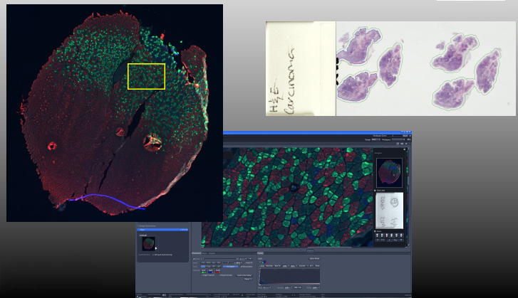 Sample images from the slide scanner. Fluorescence images show a multi-channel, stitched image (top left) with a screen shot of the zoomed-in inset (center; image courtesy of Sabine Jordan). Top right shows a preview scan of a slide with H&E-stained sections (image courtesy of Anna Robles).
How long does each slide take to scan?
It varies widely, depending on the size of your sample, the magnification you’re using, and the overall exposure time (i.e., 4-channel fluorescence will always take longer than brightfield). The good news is that it’s automated, so once you set up your scan, you can press “Start Scan” and come back when your slides are done. We even have remote desktop set up, so you can login remotely and check on your scan status.
Can someone help me with slide scanning?
Yes! We are now offering full-service slide scanning. Please fill out the User Request Form for more details.